
Conduction system of the heart and electrocardiography exercise 31 – In the realm of cardiac physiology, the conduction system of the heart and electrocardiography (ECG) stand as fundamental pillars, orchestrating the rhythmic beating of our hearts and providing invaluable insights into its electrical activity. This exploration, centered around Exercise 31, delves into the intricate mechanisms that govern the heart’s electrical impulses, their manifestation on ECG recordings, and the profound impact of exercise on these vital processes.
The electrical conduction system, a marvel of biological engineering, ensures the coordinated contraction of the heart chambers. From the sinoatrial node, the heart’s natural pacemaker, to the Purkinje fibers that distribute electrical impulses throughout the ventricles, each component plays a crucial role in maintaining a regular heartbeat.
Electrocardiography, a non-invasive technique, captures the electrical activity of the heart, providing a window into its rhythm and electrical integrity. Exercise, a potent physiological stimulus, exerts significant effects on the heart’s electrical behavior, revealing valuable information about its functional capacity.
Electrical Conduction System of the Heart

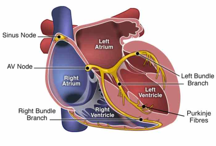
The electrical conduction system of the heart is a specialized group of cells that generate and transmit electrical impulses, coordinating the rhythmic contractions of the heart. It ensures the proper timing and sequence of electrical impulses, allowing the heart to pump blood efficiently.
Sinoatrial Node (SA Node)
The sinoatrial node (SA node) is located in the right atrium and is the natural pacemaker of the heart. It generates electrical impulses that initiate each heartbeat and sets the heart rate.
Atrioventricular Node (AV Node)
The atrioventricular node (AV node) is located between the atria and ventricles. It receives electrical impulses from the SA node and delays them slightly before transmitting them to the ventricles. This delay allows the atria to fill with blood before the ventricles contract.
Bundle of His
The bundle of His is a bundle of fibers that connects the AV node to the left and right bundle branches. It conducts electrical impulses from the AV node to the ventricles.
Bundle Branches and Purkinje Fibers
The left and right bundle branches extend from the bundle of His and distribute electrical impulses throughout the ventricles. Purkinje fibers are specialized fibers that conduct electrical impulses within the ventricular myocardium, ensuring coordinated contraction of the ventricles.
Electrocardiography (ECG): Conduction System Of The Heart And Electrocardiography Exercise 31
Electrocardiography (ECG) is a non-invasive technique that records the electrical activity of the heart. It is used to diagnose and monitor heart conditions.
Principles of Electrocardiography
An ECG records the electrical changes on the body’s surface that result from the electrical activity of the heart. Electrodes are placed on the skin at specific locations to capture these changes.
ECG Leads
Different ECG leads provide different views of the heart’s electrical activity. The most common leads are the 12-lead ECG, which includes leads from various positions on the chest, arms, and legs.
ECG Waveform Components
An ECG waveform consists of distinct waves and intervals that represent the electrical events of the heartbeat:
- P wave: Atrial depolarization
- QRS complex: Ventricular depolarization
- T wave: Ventricular repolarization
Clinical Significance of ECG Findings
ECG findings can provide valuable information about the heart’s electrical activity, including heart rate, rhythm, and any abnormalities. ECGs are used to diagnose conditions such as arrhythmias, myocardial infarction, and electrolyte imbalances.
Electrocardiography in Exercise

Exercise can significantly affect the ECG.
Effects of Exercise on the ECG
Exercise increases heart rate, stroke volume, and cardiac output. These changes are reflected on the ECG as:
- Increased heart rate
- Taller P waves
- Wider QRS complex
- Increased amplitude of the T wave
ST-Segment in Exercise ECG
The ST-segment is the portion of the ECG waveform between the QRS complex and the T wave. In exercise ECG, the ST-segment is closely monitored as it can provide information about myocardial ischemia.
Arrhythmias During Exercise
Exercise can trigger or unmask arrhythmias. Identifying and interpreting arrhythmias in exercise ECG is important for assessing cardiac health.
Clinical Applications of Exercise ECG
Exercise ECG is used to:
- Diagnose exercise-induced arrhythmias
- Assess myocardial ischemia
- Monitor the effectiveness of cardiac rehabilitation programs
Exercise Physiology
Exercise physiology studies the physiological responses to exercise.
Physiological Responses to Exercise
Exercise induces a cascade of physiological adaptations:
- Increased cardiac output
- Increased respiratory rate and depth
- Increased muscle blood flow
- Hormonal changes (e.g., increased adrenaline and growth hormone)
Cardiovascular and Respiratory Adaptations
Exercise training improves cardiovascular and respiratory fitness:
- Increased stroke volume
- Reduced heart rate at rest and during exercise
- Increased lung capacity
- Improved oxygen utilization
Hormonal Changes During Exercise
Exercise triggers hormonal changes that support energy production and recovery:
- Increased adrenaline (epinephrine): Mobilizes energy stores
- Increased growth hormone: Stimulates muscle growth and repair
- Increased cortisol: Regulates metabolism and reduces inflammation
Exercise Training Programs
Exercise training programs are designed to improve physical fitness and overall health:
- Aerobic exercise: Improves cardiovascular fitness (e.g., running, cycling)
- Strength training: Improves muscle strength and mass (e.g., weightlifting)
- Flexibility training: Improves joint range of motion (e.g., yoga, stretching)
Case Study: Analysis of an Exercise ECG
An exercise ECG can provide valuable information for diagnosing and managing cardiac conditions.
ECG Findings
In this case study, the exercise ECG showed:
- Increased heart rate (180 bpm)
- Tall P waves
- Wide QRS complex (120 ms)
- ST-segment depression in the inferior leads
Clinical Significance, Conduction system of the heart and electrocardiography exercise 31
The ECG findings suggest:
- Sinus tachycardia (rapid heart rate)
- Right ventricular hypertrophy (thickening of the right ventricular wall)
- Possible myocardial ischemia in the inferior wall of the heart
Management Plan
Based on the ECG findings, the following management plan was recommended:
- Further evaluation with echocardiography to confirm right ventricular hypertrophy
- Stress testing to assess myocardial ischemia
- Referral to a cardiologist for further evaluation and treatment
FAQ Section
What is the sinoatrial node?
The sinoatrial node is the natural pacemaker of the heart, responsible for initiating electrical impulses that trigger each heartbeat.
How does exercise affect the ECG?
Exercise increases heart rate and cardiac output, leading to changes in the ECG, including ST-segment depression or elevation, which can provide insights into myocardial ischemia or injury.
What is the clinical significance of arrhythmias during exercise?
Arrhythmias during exercise can indicate underlying heart conditions, such as coronary artery disease or electrolyte imbalances, and require further evaluation.
